Please note that some guidelines may be past their review date. The review process is currently paused. It is recommended that you also refer to more contemporaneous evidence.
Bowel obstruction is a common surgical emergency for newborns. Early diagnosis and appropriate treatment usually results in positive outcomes. Delay in carrying out surgery may result in the loss of large amounts of bowel.
Not all infants with bowel obstruction require transfer by PIPER neonatal. Infants diagnosed early and without fluid or electrolyte problems may be safely transferred with local ambulance services. However, it is advisable to discuss such infants with the receiving hospital or PIPER.
Signs of bowel obstruction
Signs of bowel obstruction can include:
- vomiting with or without bile stained material; never ignore bile-stained vomiting in the newborn
- increased gastric residuals before feedings
- failure to pass meconium in the first 24 hours of life
- abdominal distension (particularly with low level obstruction)
- absent or decreased bowel sounds.
Differential diagnosis
Intestinal obstruction without bilious vomiting
This may occur in:
- duodenal atresia (if obstruction proximal to Ampulla of Vater 20 per cent of cases)
- duodenal stenosis
- annular pancreas
- pyloric stenosis (usually presents at four to six weeks of life but may present as early as the first week).
Intestinal obstruction with bilious vomiting
This may occur in:
- malrotation and volvulus
- duodenal atresia (if obstruction distal to Ampulla of Vater 80 per cent of cases)
- jejunoileal atresia
- meconium ileus
- meconium plug syndrome (18 per cent)
- Hirschsprung’s disease (8 per cent).
Intestinal obstruction with marked abdominal distension
This may occur in:
- ileal atresia
- Hirschsprung's disease
- meconium ileus
- meconium plug syndrome
- imperforate anus.
Duodenal atresia
Duodenal atresia may take the form of either a membranous or interrupted-type lesion at the level of the papilla of Vater.
In 80 per cent of cases the papilla of Vater opens into the proximal duodenum causing the vomiting to be bilious.
Facts about duodenal atresia:
- obstruction is due to failure of recanalisation of the second part of the duodenum during fetal development
- occurs in 1:5,000-10,000 live births
- more common in males
- associated with Down syndrome in 25 per cent of cases
- polyhydramnios is seen antenatally
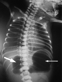
- x-ray usually shows a characteristic 'double-bubble' appearance.
 Figure 1: X-ray showing characteristic ‘double bubble’ appearance in duodenal atresia
Midgut malrotation and volvulus
Malrotation is a result of abnormal rotation of the gut as it returns to the abdominal cavity during embryogenisis. It occurs in 1:6000 live births and is often associated with other conditions including duodenal atresia or stenosis, congenital diaphragmatic hernia, abdominal wall defects, heterotaxy or choanal atresia.
Most patients with midgut malrotation develop volvulus within the first week of life. Features include the following:
- Bilious vomiting is the initial symptom and abdominal distention is minimal until a much later stage.
- Bowel can become strangulated at any time and age. Once midgut ischaemia occurs, unstable haemodynamics, intractable metabolic acidosis and necrosis with perforation develop.
- Abdominal x-ray may be normal.
- Malposition of the superior mesenteric vessels demonstrated by ultrasound examination is diagnostic.
- Upper gastrointestinal contrast study is the investigation of choice but should be performed by experienced practitioners only. The key feature with malrotation is:
- abnormal position of the duodeno-jejunal (DJ) junction (the DJ junction fails to cross to the left of the vertebral bodies and lies inferior to the first part of the duodenum)
- spiral configuration of the jejunum or a duodenojejunum that occupies the right hemi-abdomen is seen with volvulus.
- Symptomatic infants require immediate surgery.
Jejunoileal atresia
Jejunoileal atresia is caused by a mesenteric vascular accident during fetal life. Features include the following:
- Abdominal distention with bilious vomiting is observed within the first 24 hours after birth. The more proximal the lesion, the earlier the bile-stained vomiting.
- X-ray shows air-fluid levels proximal to the lesion.
- Calcification due to meconium peritonitis may be present.
Meconium ileus
Thick tenacious meconium in the bowel (ileum, jejunum or colon) causes obstruction. Fifty per cent of infants may have associated:
- volvulus
- jejunoileal atresia
- bowel perforation and/or meconium peritonitis.
Meconium ileus occurs in 15 per cent of newborns with cystic fibrosis, and at least 90 per cent of patients with meconium ileus have cystic fibrosis.
Presentation of meconium ileus
Presentation includes:
- early marked bowel distension
- bilious vomiting
- remarkable abdominal distention, tenderness and/or erythema of the abdominal skin may indicate perforation
- on rectal examination mucus plugs may be evacuated after withdrawal of the examination finger (fifth finger).
X-ray investigation of meconium ileus
X-ray shows:
- distended loops of intestine with thickened bowel walls
- a large amount of meconium mixed with swallowed air produces the so-called 'ground-glass' sign typical of meconium ileus, a characteristic feature but often absent
- calcification, free air or very large air-fluid levels suggest bowel perforation.
Treatment of meconium ileus
Patients with uncomplicated meconium ileus may be successfully treated with hypertonic enemas performed while adequate intravenous fluid is maintained.
Immediate surgery is indicated for infants with complicated meconium ileus or where conservative treatment fails.
Meconium plug syndrome
- Most common form of functional distal intestinal obstruction in the neonate
- Caused by inspissated meconium within the distal colon or rectum
- Occurs in 1:500-1:1000 live births, with uncertain etiology
- Generally present with marked abdominal distension and failure to pass meconium
- Generalised intestinal dilatation is seen on x-ray
- Contrast enema is diagnostic showing outlines of the meconium plugs
- Contrast enema or digital exam is often curative with the plugs being extruded
- Stooling pattern should be normal afterwards
- If any concern about stooling pattern a rectal suction biopsy should be performed to rule out Hirschsprung’s disease
Hirschsprung's disease
Hirschsprung's disease is the cause of 15-20 per cent of newborn intestinal obstructions occurring in 1:4000 live births. It is characterised by abnormal innervation of the colon. It can affect the anal sphincter or extend throughout the entire colon into the small bowel.
Features:
- 80 per cent of cases present in the first six weeks of life.
- 4:1 male:female ratio.
- 8 per cent of patients also have Down syndrome
- Presents with failure to pass meconium in the first 24 hours plus gradual onset of abdominal distension and vomiting.
- Distal short segment disease can present later in life with persistent and progressive constipation.
- The most serious complication is enterocolitis. This occurs as a result of progressive colonic dilation with decreased ileal and colonic fluid resorption, stasis with bacterial overgrowth and mucosal ischaemia, which may lead to massive acute fluid loss into the bowel with diarrhoea, shock and dehydration.
- Enemas should be avoided during episodes of enterocolitis because of the possibility of perforating the colon.
Diagnosis of Hirschsprung's disease
A definitive diagnosis is made by a suction rectal biopsy with acetylcholinesterase staining showing prominent nerve fibres and absence of ganglion cells in the submucosa. There is absence of ganglion cells in the myenteric plexus of the colon on histological examination.
Investigation of bowel obstruction
When you investigate bowel obstruction you should:
- Conduct a thorough physical examination and an assessment of the circulation with documentation of findings.
- Look for any associated abnormalities because bowel obstruction may be a part of multiple anomalies, for example: vertebral, anal, cardiac, tracheo-esopageal atresia, renal, limb (VACTERL) sequence with imperforate anus, trisomy 21 with duodenal atresia.
- Get plain abdominal x-rays.
- Note that digital rectal examination, contrast studies and ultrasound examinations are best undertaken in centres with paediatric surgical services
Management of bowel obstruction
To manage bowel obstruction you should:
- Place the infant in an incubator for close observation and temperature control.
- Nurse supine with the head elevated.
- Place an orogastric tube (8-10FG) on low-pressure suction (or aspirate with a syringe every 60 minutes and leave on free drainage). The amount and type (for example, bile-stained, faeculent) of fluid aspirated should be recorded.
- Place nil by mouth.
- Commence IV fluids. If signs of shock may need fluid resuscitation with normal saline in 10-20 mL/kg aliquots. Give maintenance fluids plus mL for mL replacement of NG aspirate with normal saline and 10 mmol KCL/500 mL.
- Obtain abdominal x-rays (include supine and lateral decubitus view).
- Note that a relatively gasless abdomen is compatible with mid-gut volvulus.
- Consult with a paediatric surgeon or PIPER neonatal to arrange transfer to an appropriate surgical centre.
- It may be appropriate to commence antibiotics preferably after blood culture taken (discuss with the receiving unit or PIPER).
- Obtain blood for FBE, electrolytes, blood gas and lactate (and blood cultures if commencing antibiotics).
- Be aware that these infants frequently have associated problems of acidosis and shock.
More information
References
Hutson, J. et al (eds) Jones Clinical Paediatric Surgery Diagnosis and Management, 6th ed., 2008, Blackwell Publishing
Get in touch
Version history
First published: September 2013
Due for review: September 2016
