Please note that some guidelines may be past their review date. The review process is currently paused. It is recommended that you also refer to more contemporaneous evidence.
Pneumothorax drainage topic includes clinical features of pneumothorax, preparation for procedure, emergency needle aspiration and procedure for insertion of an intercostal catheter.
Pneumothorax occurs when air escapes from ruptured alveoli into the pleural cavity ( the potential space between the lung and the chest wall). Whereas a small collection of air may not compromise the infant, accumulation of larger air volumes may result in collapse of the ipsilateral lung and shift of the mediastinum to the contralateral side. When the accumulating air is under pressure a tension pneumothorax results. This is an acutely life threatening situation and immediate drainage will be required. In the acute situation needle aspiration is performed, followed by intercostal catheter (ICC) insertion.
A pneumothorax diagnosed as an incidental finding on chest x-ray may not require active drainage, but when associated with clinical deterioration, it may require expedient drainage. The incidence of pneumothorax is dramatically lower since the advent of surfactant and with continuous positive airway pressure (CPAP) rather than ventilation, but air leaks still occur in 5-10 per cent of babies with neonatal lung disease.
Clinical features of pneumothorax
Pneumothorax should be considered with:
- sudden deterioration with oxygen desaturation/increased oxygen requirement
- increase in respiratory distress and/or diminished chest movement
- circulatory compromise (indicates mediastinal shift/compression)
- hypoxia, respiratory and/or metabolic acidosis on blood gas.
Clinical signs include:
- decreased air entry and hyperresonance on percussion of the affected side
- abdominal distention due to displacement of the diaphragm
- displaced apex beat
- hyperluminescence with transillumination.
These signs are, however, unreliable in:
- infants with increased thickness of the chest wall, for example, term infants and oedema
- infants with pulmonary interstitial emphysema (who may show a 'false positive' result).
Chest x-ray will confirm the diagnosis but takes time to perform.
If a tension pneumothorax is suspected clinically, immediate aspiration should not be delayed to obtain an x-ray.
Preparation
Infants breathing spontaneously should be monitored to determine if they need intubation and ventilation.
Consider the requirement for appropriate pain relief. This may include:
- oral sucrose
- infiltration of the insertion site with 1 per cent lignocaine 0.5 -1 mL before preparing and draping the field (in order to allow greater time for the anesthetic to take effect)
- intravenous infusion of morphine.
Needle aspiration emergency option
Needle aspiration is an emergency procedure only.
Equipment
- Butterfly needle
- 25G for < 32 w or < 1,500 g
- 23G for > 32 w or > 1,500 g
- 3-way stopcock
- 10 mL syringe
- 70 per cent isopropyl alcohol swab
- one pair sterile gloves.
 Figure 1: needle for emergency drainage
Procedure
- Position infant supine, prepare area with alcohol wipe
- Insert needle into the pleural space (directly over the top of the rib in the second or third intercostal space in the midclavicular line) until air is aspirated into the syringe. Expel air through the three-way stopcock.
- Minimise movement in the needle to avoid lacerating the lung or puncturing blood vessels.
 Figure 2: Position for needle aspiration
Ongoing care
Following needle aspiration, insertion of an intercostal catheter is required for ongoing management. It may be necessary to seek help with this procedure - consultation and assistance will be available through PIPER or the receiving NICU.
Drainage
The preferred drain is a Fuhrman pigtail catheter, but the alternative remains a trocar catheter. The insertion procedure will be described for both. Once the catheter has been inserted it is immediately connected to either a one way valve (Heimlich valve) or an underwater seal drainage system (with or without active suction).
Intercostal catheters can also be used to drain pleural effusions.
Equipment for pigtail catheter insertion
- 15 cm long polyurethane Pigtail catheter with 6 side ports
- 10 Fr. for > 1,500 g
- 8 Fr. for < 1,500 g
- Sterile introducer needle, guidewire, dilator and connector tubing and three-way tap as packed by supplier
- 1 per cent lignocaine syringe and needle
- Skin preparation
- Underwater seal drainage system or Heimlich valve
- Sterile gown, gloves and drapes
- Semi-occlusive dressing, tapes
 Figure 3: Equipment for pigtail catheter
Procedure for intercostal catheter insertion
- Observe standard precautions.
- Mask, sterile gown and gloves are required as for any sterile procedure.
- Place infant under radiant heater to maintain infant's temperature.
- Monitor heart rate and saturation levels and ensure infant can still be partly visualised after draping to create a sterile field.
- Position the infant with the effected side uppermost and the arm extended above the head (a nappy cloth roll may help maintain a good position). Ensure limbs are adequately restrained.
- Monitor infant's heart rate and oxygen saturation level.
- Prepare the field with antiseptic solution and drape.
- Infiltrate local anaesthetic at insertion site (fourth or fifth intercostal space in the anterior axillary line. This corresponds to a point 1-2 cm lateral to and 0.5-1 cm below the nipple).
- Mark off 1.5 cm on the introducer needle with a steri-strip or place a clamp in this position. Connect the needle to a small syringe with a small amount of sterile water (to see air bubbles whilst aspirating).
- Advance the needle through the infiltrated skin, gently aspirating until air is obtained. Continue to aspirate if pneumothrorax is under tension.
- Remove syringe, occlude temporarily, then thread the guidewire through the hub of the insertion needle via the white plastic tip (fits nicely into the hub and straightens out the curved tip of the guidewire). Advance until the silver guideline on the wire reaches the white plastic tip.
- Remove the needle while not allowing the wire to move (clamp the wire at the skin as soon as the needle is out of the way).
- Thread the dilator over the guidewire and insert about 1 cm through the skin withdraw and remove the dilator.
- Feed pigtail catheter over the guidewire with the holes facing up. Advance to first to second black line for a premature infant, fourth to fifth for a term infant.
- Remove the guidewire.
- Connect the catheter to the connection tubing via the tap. The other end of the tubing connects to the Heimlich valve or the underwater drainage system. Note whether the fluid is swinging and/or bubbling. Fogging within the catheter may be seen when within the pleural space.
- Secure the pigtail with a steristrip (Roman sandal around) and then Tegaderm.
Ongoing care
- Check the tube position and resolution of the pneumothorax by transillumination and x-ray as soon as possible.
- Determine the need for ongoing analgesia based on an assessment of physiological and behavioural responses associated with pain.
- Transfer infants who require an intercostal catheter to an NICU if required for ongoing care.
Equipment Trocar intercostal catheter insertion
- Argyle 8, 10 or 12 Fr sterile intercostal catheter
- Sterile surgical instrument pack
- Size 11 scalpel blade
- 3/0 black silk suture on a curved edge needle
- Skin preparation
- 1 per cent lignocaine, syringe and needle
- Underwater seal drainage system or a Heimlich valve
- Sterile gown, gloves and drapes
- Semi-occlusive dressing, tapes
Procedure Trocar intercostal catheter insertion
- Observe standard precautions.
- Mask, sterile gown and gloves are required as for any sterile procedure.
- Place infant under radiant heater to maintain infant's temperature.
- Monitor heart rate and saturation levels and ensure infant can still be partly visualised after draping to create a sterile field.
- Position the infant with the effected side uppermost and the arm extended above the head (a nappy cloth roll may help maintain a good position). Ensure limbs are adequately restrained.
- Prepare the field with antiseptic solution and drape.
- Infiltrate local anaesthetic at insertion site (fourth or fifth intercostal space in the anterior axillary line. This corresponds to a point 1-2 cm lateral to and 0.5-1 cm below the nipple).
- Select intercostal catheter size:
|
Infants |
> 1,500 g |
10 or 12 Fr |
|
|
< 1,500 g |
8 or 10 Fr |
Make a 1 cm incision through the skin and subcutaneous tissue using a small (number 11) scalpel blade.
Bluntly dissect away the subcutaneous tissue and intercostal muscles using straight mosquito forceps to reach the parietal pleura. Aim to dissect a passage just above a rib border in order to avoid the neurovascular bundles running below each rib. Open the parietal pleura by blunt dissection. At this point the hiss of air escaping the pleural space may be heard.
Remove the trocar from the ICC and grasp the distal end with curved artery forceps. Advance the ICC into the pleural space 3-5 cm (at the 1-3 cm marking on the catheter), directing the tip anteriorly as well as superomedially, so that the tip lies anteriorly inside the chest cavity.
Connect the ICC to a Heimlich valve or an underwater seal drainage system, and note whether the fluid is swinging and/or bubbling. Fogging within the catheter may be seen when within the pleural space.
Place a single stitch through the wound so that the skin is drawn snugly around the ICC. Purse string stitches are not used as they leave an unsightly scar. Wrap the ends of the suture around the ICC several times and tie securely.
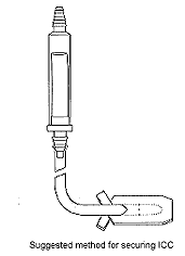
Secure the ICC to the chest wall with trouser leg tapes as shown in diagram. This helps to maintain the anterior position of the ICC and minimises trauma to intrathoracic structures due to movement of the extrathoracic portion of the ICC.
Alternatively, sandwich the wound and tube between two Tegaderm dressings.
 Figure 4: Diagram for securing ICC
Ongoing care
- Check the tube position and resolution of the pneumothorax by transillumination and x-ray as soon as possible.
- Determine the need for ongoing analgesia based on an assessment of physiological and behavioural responses associated with pain.
- Transfer infants who require an intercostal catheter to an NICU if required for ongoing care.
More information
References
- Stabilisation and Transport of Newborn Infants and At-Risk Pregnancies. Editors ED Bowman, SM Levi, FE Presbury, A McLean. Newborn Emergency Transport Service, 4th edition, 1998.
- Intercostal catheters in Neonates- Insertion & care. Clinical protocols & guidelines, Southern Health
Get in touch
Version history
First published: March 2014
Review by: March 2017

