Acknowledgement
This guidance uses the terms ‘woman’ and ‘mother,’ which are intended to be inclusive of anyone who may use other self-identifying terms and aims to encompass all for whom this guidance is relevant.
Consumer Engagement Statement
All interactions between health care staff with consumers (women, mothers, patients, carers and families) should be undertaken with respect, dignity, empathy, honesty and compassion.
Health care staff should actively seek and support consumer participation and collaboration to empower them as equal partners in their care.
Definitions/Abbreviations
| MFM | Maternal Fetal Medicine |
|---|---|
| NGT | Nasogastric tube |
| NICU | Neonatal Intensive Care Unit |
| PIPER | Paediatric, Infant Perinatal Emergency Retrieval |
Introduction
This guideline will discuss the management principles of the two common abdominal wall defects: gastroschisis and exomphalos.
While parts of this guideline are written for Victorian capability level 6 services, the resuscitation, stabilisation and transfer sections are relevant across all levels of health service capability.
In all cases of gastroschisis and especially in unexpected births occurring outside of capability level 6 hospitals, the management priorities are as detailed in the Immediate post-birth management of abdominal wall defects section.
Notify PIPER (1300 137 650) when the neonate is born (do not wait until stabilisation is complete).
Gastroschisis is characterised by the herniation of the neonate's bowel and other abdominal contents through an abdominal wall defect, usually located just to the right of the umbilical cord.
The normal insertion of the umbilical cord and the absence of a sac covering the herniated abdominal contents distinguishes gastroschisis from exomphalos (the other common abdominal wall defect). The defect likely occurs between the fourth and tenth week of gestation. Gastroschisis occurs in approximately 1 in 4000 births in Victoria. 1
An exomphalos (also known as omphalocele) is the herniation of the neonate’s abdominal organs through a central abdominal wall defect. A sac comprised of peritoneum, amnion, and Wharton’s jelly covers the central herniation into the umbilical cord, whereas there is no sac protecting the exposed bowel in gastroschisis.
Exomphalos occurs when intestines fail to return to the abdominal cavity after the normal embryonic herniation into the umbilical cord between the sixth and tenth week of gestation. 2
Exomphalos occurs about 1 in every 2500 births in Victoria. An exomphalos is more likely to have associated major congenital anomalies or to be part of a syndrome. 1
There are two types of exomphalos:
- Exomphalos minor where the opening is less than four centimetres and only contains the intestine and omentum.
- Exomphalos major (or Giant exomphalos) where the opening is greater than four centimetres and may contain liver as well as intestine.
In Victoria, approximately ten neonates are born each year with this condition and, on average, three of them have the major form of the anomaly.
Management
The following document provides guidance for the management of abdominal wall defects from antenatal diagnosis to post-operative and continuing care.
Antenatal
More than 90 per cent of cases of gastroschisis are antenatally diagnosed and almost 100% of cases of exomphalos are diagnosed antenatally via ultrasound. 2
In Victoria, pregnancies with an antenatal diagnosis of abdominal wall defect are managed at a capability level 6 perinatal centre in the Maternal Fetal Medicine Unit.
Parents of neonates that are antenatally diagnosed should receive multidisciplinary counselling in the antenatal period, including consultant paediatric surgical input.
It is important to ensure the expectant parents feel supported and able to ask questions. They need to be prepared for how their neonate will look after birth and understand the anticipated treatment plan. Ideally families should be offered a broad orientation to the NICU environment antenatally. Familiarity with the NICU environment will assist parents to be as prepared as possible for their neonate's admission.
Other conditions associated with abdominal wall defects
Gastroschisis is usually an isolated problem, rarely associated with chromosomal abnormalities or other major non-gastro-intestinal congenital abnormalities. Between 10-15% have associated intestinal atresia. 6
In contrast, associated anomalies are observed in up to 72% of neonates with exomphalos. 3
Of neonates with normal karyotypes, nearly 80% have multiple other complications or anomalies. Multiple anomalies are more common with minor (< 4 cm) versus major exomphalos (55% vs 36%). 3
Table 1. Exomphalos associations
| Chromosomal abnormalities | Trisomy 13, 14, 15, 18 and 21 |
|---|---|
| Syndromes | Beckwith-Wiedemann Syndrome, Pentalogy of Cantrell, Lower Midline Syndrome (bladder/cloacal extrophy, anorectal malformation, myelomeningocele), Donnai-Barrow Syndrome |
| Cardiac anomalies | Seen in up to 20% of cases |
| VACTERL association | Exomphalos may occur in association with Vertebral, Anorectal, Cardiac, Tracheo-oesophageal fistula and Oesophageal Atresia, Renal Tract and Limb anomalies |
| Pulmonary hypoplasia | Commonly associated with exomphalos major (20%) |
| Intestinal Abnormalities | A rotational anomaly is nearly universal; intestinal atresia’s are found in <10% |
| Nervous system | Holoprosencephaly and anencephaly |
| Other | Cleft lip and palate, polydactyly, limb reduction defects |
Women should be referred to a multidisciplinary fetal medicine service as soon as possible after diagnosis, so that detailed testing may be arranged to assess for the presence of the other known associations (as above) and options for ongoing care and management pathways should be discussed.
Birth should be planned at a capability level 6 maternity service and the anticipated accepting surgical NICU team should be integral in development of the management plan and aware of the anticipated date of birth.
If birth (via either caesarean or induction of labour) is planned, it is important that the perinatal team notify the anticipated accepting surgical NICU to confirm bed availability before commencing the birth process. If there is an imperative to urgently deliver the neonate, NICU access will not influence the timing of such births.
Mode of birth
For neonates with uncomplicated gastroschisis and in the absence of other risk factors, vaginal birth may be safe. 4 Induction or semi-elective caesarean section is often necessary due to concern about fetal wellbeing, and the risks associated with the exposed bowel. There is a significant incidence of late preterm birth due to concerns for fetal well-being and integrity of exposed bowel. In such circumstances, Caesarean section may be advised. The median gestation at birth is 36 weeks.
In exomphalos major, vaginal birth risks liver injury or sac rupture. Birth by caesarean section is advised to minimise these risks. Subsequent management of the neonate is then dictated by the size of the exomphalos, associated anomalies and whether the sac is intact after birth. 5
Immediate post birth management of abdominal wall defects
Prepare as for anticipated high-risk birth, as per unit protocol. Senior Medical staff should attend the birth.
In the rare event that a neonate with unanticipated gastroschisis or exomphalos births outside of a level 6 capability service, all birthing units should have a roll of cling wrap readily available and clinicians should follow instructions below for immediate management.
Manage airway, breathing and cardiovascular status as per usual practice. In the compromised neonate endotracheal intubation should be undertaken early to minimise gut distension. Prolonged mask ventilation or non-invasive ventilation should be avoided for this reason.
In exomphalos, some neonates may have unsuspected pulmonary hypoplasia. Congenital heart disease should be suspected if the neonate is cyanosed/not responding to resuscitation.
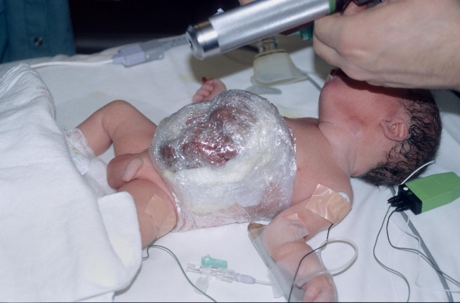
Once the cardiorespiratory status has been stabilised, quickly inspect the bowel, correcting any obvious twists on its pedicle. The bowel should then be positioned centrally over the abdomen, supported and wrapped in cling wrap as described below.
Insert an 8Fr nasogastric tube and aspirate the stomach, then leave on free drainage. Ensure adequate thermal control.
Immediate management of gastroschisis
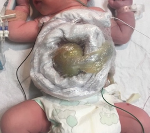
Following assessment, the exposed bowel should be wrapped with cling wrap (transparent, latex free) for protection and to minimise fluid and heat loss: see Figure 1.

Figure 1
This is achieved by:
- A clean procedure that does not need to be sterile.
- Slide large piece of cling wrap under the neonate’s buttocks and back with a length of wrap extending either side of the neonate.
- Place exposed organs on neonate’s abdomen (using sterile latex-free gloves).
- Wrap cling wrap gently around the abdomen and exposed organ.
- Ensure that the bowel is not exposed to drying air.
- Avoid compressing the bowel, it should remain mobile but entirely covered.
- Monitor the bowel by visual inspection through the transparent wrap every 15 minutes for dusky or blanching colour changes.
- Remove and rewrap as above if compression, kinking or twisting of bowel is suspected and If there is no suspicion of increased bowel compromise, unwrapping and handling of the bowel should be strictly avoided.
- Support the intestines to prevent occlusion of the blood supply where the bowel exits the defect in the abdominal wall.
- If necessary, support the exposed intestines with your hands and where possible, nurse the neonate on their right side, with the wrapped bowel supported perpendicular to the umbilicus using a rolled towel or equivalent.
Immediate management of exomphalos
| Exomphalos with intact sac | Exomphalos with ruptured sac |
|---|---|
|
|

Figure 2: Exomphalos doughnut (Source: Royal Women's Hospital)
Perinatal Unit
General
The objective in all babies with gastroschisis is to stabilise and transfer as soon as possible, within four hours of birth.
- The urgency of transfer to a surgical NICU in exomphalos depends on the size of defect and presence of associated anomalies.
- Notify PIPER (1300 137 650) when the neonate is born (do not wait until stabilisation is complete).
- The principles of management following birth and during the pre-transfer period includes addressing fluid resuscitation, gastric decompression, avoidance of hypothermia and care of the herniated abdominal contents.
- Manage the airway, breathing and any cardiovascular instability as per standard practice.
- Ensure the protruded abdominal contents remain wrapped and supported as per the methods described above.
- Ensure NGT is aspirated hourly and remains on free drainage.
- Establish vascular access. The minimum required will be two peripheral intravenous cannulas. Arterial access is not required at this stage unless there is significant respiratory or circulatory compromise. Collect blood sample for baseline gas, electrolytes, glucose and culture when IV inserted.
- Give intravenous benzylpenicillin and gentamicin (preferably after blood culture).
- Check blood glucose immediately and monitor closely because of the association of Beckwith-Wiedemann syndrome with exomphalos minor.
- Ensure the neonate is clinically assessed for other congenital anomalies.
- Ensure continued thermal control. This needs to be closely monitored due to increased evaporative heat loss.
- Ensure appropriate cardiorespiratory and saturation monitoring is applied and close monitoring of blood pressure occurs.
Fluids
- Insensible losses will be unavoidably high. Newborns with abdominal wall defects require up to 2-3 times the amount of fluid of a normal term newborn and thus fluid resuscitation is required early.
- Commence maintenance fluid 10% Dextrose at 60 ml/kg/day to maintain blood glucose >2.6 mmol/L.
- Give a 20 ml/kg fluid bolus of normal saline within an hour of birth.
- Run an additional 10 ml/kg/hr of normal saline as the default minimum ongoing replacement fluid.
- Maintain an accurate fluid balance record, including gastric losses.
- Regular assessment of overall fluid and clinical status should occur with a low threshold for repeated normal saline bolus.
- Once 40mL/kg of normal saline has been administered, ongoing fluid replacement needs to take account of possible clotting factor dilution and/or the need for colloid/blood products as opposed to further normal saline. 6
Surgical considerations
- The paediatric surgical team should be notified of the neonate’s birth. Surgical review should occur as soon as possible after arrival in the surgical NICU. Operation to close the abdominal wall defect is usually planned within four hours of arrival in the surgical NICU. Management of an associated atresia is generally delayed until a subsequent surgery.
- In more complex cases of gastroschisis (perforation, atretic segments) a 'clip and drop' of the discontinuous bowel approach may be required with definitive management delayed to a subsequent surgery when the neonate is better able to tolerate a prolonged procedure.
- In exomphalos with intact sac, in consultation with the treating surgical team, transfer and surgery may be able to be performed electively. If the sac is ruptured, transfer and urgent surgical referral is required. Treatment of exomphalos is individualised and depends on the size of the defect, gestational age and presence of associated anomalies. Small defects may be repaired with excision of the sac and primary closure of the fascia and skin.7
- In larger exomphalos defects:
- Primary closure may be difficult due to an excessively high intra-abdominal pressure if all the contents are pushed back into the abdominal cavity.
- In these cases, the sac is left intact and allowed to slowly granulate and eventually epithelialise over several months or even years.
- Neonates do not need to remain in hospital for the duration of the epithelialisation process and may be discharged home if they are not requiring respiratory support once they are able to consume sufficient calories to grow.
- Removal of the epithelialised sac, also known as the shell, and final closure of the fascia and skin will be undertaken by the surgical team when the sac contents have returned to the abdominal cavity. 7
Surgical complications
- Short-term complications depend on the size of the lesion, type of closure, gestational age and associated anomalies. 3 These include:
- Damage to the bladder may occur if it is within the exomphalos.
- Infection risk.
- Abdominal compartment syndrome in primary closure.
- Bowel adhesions may develop post-operatively.
- Volvulus of a mal-rotated bowel.
- Short gut syndrome if extensive resection is required. 3
Other complications
- Gastro-oesophageal reflux (GOR) is common.
- Volvulus may occur, as all neonates with exomphalos have non-rotation of their intestine
- Ventral and inguinal herniae. 2
Post-operative considerations
- Feeds will start when the surgical and neonatal teams agree the neonate is systemically well and gastrointestinal function is normalising, as evidenced by passage of flatus and/or stool and reduced fluid losses via the oro/nasogastric tube.
- Time to reach full enteral feeds varies and can take several months, with the utilisation of parenteral nutrition in the interim.
- Some neonates with abdominal wall defects may require oro/nasogastric tube feeds post hospital discharge. Careful discharge planning is required to ensure that regular review for growth assessments and changing of any ongoing dressings are arranged.
Additional non-acute considerations
- Anomaly screening
- Cranial, cardiac and renal ultrasound scans are performed if there are specific clinical indicators.
- Counselling should be provided to parents when results of assessments for other anomalies are available and should take the neonate’s current clinical condition into account.
- Microarray may be offered prenatally in some cases. It is not usually required postnatally unless significant non-gastrointestinal anomalies are found.
- Consider discussion with a Paediatric Geneticist regarding additional methylation testing if Beckwith Wiedemann Syndrome is suspected. These children will require ongoing follow up including screening abdominal ultrasounds and alpha-fetoprotein levels due to risk of developing Wilms tumour and hepatoblastoma. 8
References
- Abdominal birth defects - Better Health Channel [Internet]. Betterhealth.vic.gov.au. 2022 [cited 2022 Jun 10]. Page reviewed on 2015 May 31. Available from: https://www.betterhealth.vic.gov.au/health/conditionsandtreatments/abdominal-birth-defects
- Christison-Lagay E, Kelleher C, Langer J. Neonatal abdominal wall defects. Semin Fetal Neonatal Med. 2011;16(3):164-72.
- Poddar R, Hartley L. Exomphalos and gastroschisis. Contin Educ Anaesth Crit Care Pain. 2009 Mar 4;9(2):48-51. doi: 10.1093/bjaceaccp/mkp001. Available from: https://www.sciencedirect.com/science/article/pii/S1743181617303189?via%3Dihub
- Bhat V, Moront M, Bhandari V. Gastroschisis: A state-of-the-art review. Children (Basel). 2020 Dec 17;7(12):302. doi: 10.3390/children7120302. PMID: 33348575; PMCID: PMC7765881
- Bruch S, Langer J. Omphalocoele and Gastroschisis. In: Purri P, editor. Newborn Surgery. 3rd ed. London: Hodder and Stoughton Ltd; 2011. p. 605-11.
- Starship Child Health-Newborn services clinical practice committee. Surgery-management of abdominal wall defects in the neonate [Internet]. Auckland: Starship Child Health; 2005 [updated April 2005; cited 2019 Aug]. Available from: https://www.starship.org.nz/guidelines/surgery-management-of-abdominal-wall-defects-in-the-neonate
- Hutson JM, O'Brien M, Beasley SW, Teague WJ, King S, editors. Jones' clinical paediatric surgery. John Wiley & Sons Incorporated; 2015.
- Kliegman R, Nelson W. Nelson textbook of pediatrics. 21st ed. Philadelphia: Saunders; 2020.
Citation
To cite this document use: Safer Care Victoria. Abdominal wall defects [Internet]. Victoria: Neonatal eHandbook; 2024 [cited xxxx] Available from: https://www.safercare.vic.gov.au/clinical-guidance/neonatal
Download this guidance
Version history
First published: February 2018
Reviewed: October 2024
Uncontrolled when downloaded
Get in touch
Safer Care Victoria

